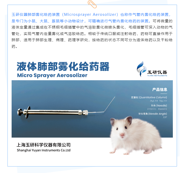


Literature Express | Yuyan Pulmonary Atomizer Device (IF=11.5) Helps Nanomedicines Reverse Pulmonary Fibrosis!
Date:2024-04-16
Author:Yuyan Instrument
Idiopathic pulmonary fibrosis (IPF) is a progressive lung disease with a high mortality rate. FDA-approved drugs, nintedanib and pirfenidone (PFD), can slow disease progression by inhibiting the overactivation of fibroblasts. However, conventional drug delivery methods result in low drug accumulation at the target site and a short duration of action, failing to significantly improve patient survival. Therefore, the search for new treatment approaches is crucial, and the emergence of nanomedicines is changing this.
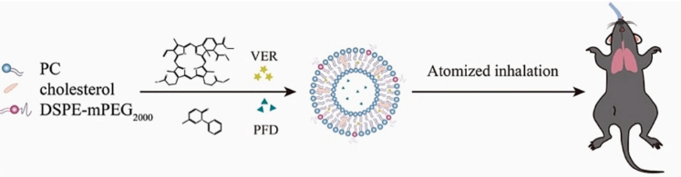
Professor Jiang Hulin's team at the School of Pharmacy at China Pharmaceutical University recently developed dual-drug loaded nanoparticles and used them with a pulmonary aerosol delivery device from Yuyan Instruments to treat pulmonary fibrosis in mice. The nanoparticles, Lip@VP, were prepared using a thin film dispersion method to simultaneously encapsulate verteporfin (VER) and pirfenidone (PFD) within a lipid layer. Using a pulmonary liquid metered dose nebulizer, a fixed amount of the nanoparticle solution was aerosolized into an aerosol with a diameter of 10-30 μm, which diffused throughout the lungs of the mice. After two weeks of treatment, the mice showed a remarkable recovery in both lung function and tissue structure. The study, titled "Inhaled nanoparticles for treating idiopathic pulmonary fibrosis by inhibiting honeycomb cyst and alveoli interstitium remodeling," was subsequently published in the Journal of Controlled Release, a leading journal in the field of drug delivery.
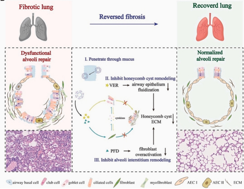
Numerous studies have reported that IPF can be alleviated by repairing damaged type II alveolar epithelial cells and inhibiting overactivated fibroblasts and dysregulated immune cells. However, further understanding has revealed that the pathogenesis of IPF is not limited to disease-specific type II alveolar epithelial cells and fibroblasts. It is well known that the airway epithelium serves as an interface between the external environment and the host. When the airway epithelium is stimulated by immune or growth factors, Yes-associated protein (YAP) is activated, driving the expression of genes associated with migration and proliferation. The effluxed cells relocate to the periphery, characterized by the proliferation and differentiation of mucin-producing airway cells. Honeycomb cysts develop in the parenchyma, and lung function declines significantly. Extensive evidence indicates that abnormal airway epithelial responses play a crucial role in disease progression.
Schematic diagram of the dual nanomedicine structure
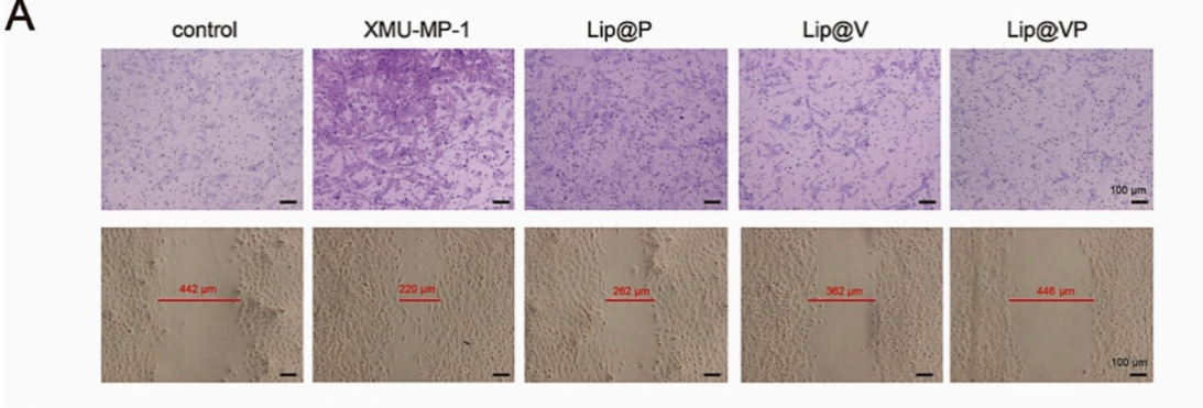
First, the authors conducted a large number of in vitro experiments, including scratch experiments to test the drug's effect on inhibiting cell migration, as well as related experiments on inhibiting cell proliferation and the drug's cytotoxicity.
The results showed that Lip@VP could effectively inhibit the migration of 16 HBE cells and the proliferation of fibroblasts.
16 HBE cell scratch assay
To verify its in vivo effects, the researchers divided mice into six groups: one group served as a normal control, and the other five groups used bleomycin (BLM) pulmonary aerosol administration to create an IPF model. Using a pulmonary aerosol drug delivery device to create a pulmonary fibrosis model has significant advantages over traditional instillation methods: pulmonary aerosolization does not require tracheotomy and there is no risk of suffocation by large droplets, so the mortality rate is significantly lower than that of the instillation method; the aerosol diffusion range is large, the drug solution is evenly distributed in the lungs, and diffuse lung lesions are closer to the actual lung condition of clinical patients; quantitative administration is possible, there is no first-pass elimination, and the drug solution is economical and highly reproducible. After 7 days of BLM treatment, the mice received aerosol administration with normal saline, Lip@P, Lip@V, and Lip@VP. At the same time, a group of mice were given oral PFD, and their lung function and pathological changes were monitored.
Schematic diagram of the experimental process
Lung function charts showed that the use of dual-drug nanoparticles (Lip@VP) could significantly improve the lung function of fibrosis mice, which was closer to that of normal control mice than other groups of mice.
Lung function indicators of mice in each group
The tidal volume, minute ventilation, peak inspiratory flow rate and mid-respiratory flow rate of each group of mice were measured by plethysmography. Compared with the normal group, the other four groups showed a significant decrease, while the Lip@VP group remained almost consistent with the normal group.
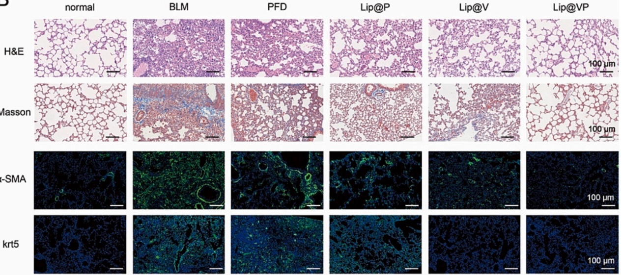
The researchers then sectioned and stained the lungs of each group of mice, performing immunofluorescence staining for α-smooth muscle actin (α-SMA) and interstitial protein (krt5). As shown in the figure below:
Lung staining of mice in each group
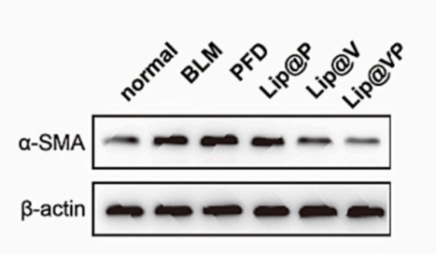
The image shows that HE staining of the lungs of normal mice reveals intact alveolar structure and a low cell count. However, in mice with fibrosis, the cell count increased significantly, and the alveolar structure became disrupted. After Lip@VP was applied to mice with pulmonary fibrosis, the alveolar structure returned to near-normal. Masson staining revealed significantly darker collagen fibers in fibrotic lung tissue, indicating that the increased number of cells is actually overproliferating fibroblasts. The expression of α-SMA and krt5 was also significantly increased in the lungs of fibrotic mice. Excessive increases in these proteins exacerbate fibrosis, while the Lip@VP group reversed this expression back to normal levels. Quantitative measurements further confirmed that the nanodrug can reduce the overexpression of α-SMA and krt5.
α-SMA and krt5 protein expression bands in each group
The study, through the modeling of pulmonary fibrosis and the reversal of fibrosis with nanomedicines, suggests that pulmonary fibrosis is a process of airway epithelial fluidization and excessive proliferation of fibroblasts. Therefore, inhibiting airway fluidization and fibroblast proliferation is the starting point for treatment. Nanomedicines enter the alveolar surface through intrapulmonary atomization due to their stronger permeability and sustained release ability; VER inhibits airway epithelial fluidization and reduces pathological cell migration, while PFD inhibits excessive fibroblast proliferation. The two work together to reshape the honeycomb cysts and alveolar distortions formed by pulmonary fibrosis, achieving a cure. Overall, the nanoparticle-loaded dual-drug experiment achieved surprising results. By encapsulating the drug in nanoparticles, its penetration and accumulation capacity are enhanced, and by pulmonary atomization, its range of action is increased, finding a new strategy for the clinical treatment of IPF.
Next is an introduction to pulmonary drug delivery.
Professor Jiang Hulin's team at the School of Pharmacy at China Pharmaceutical University recently developed dual-drug loaded nanoparticles and used them with a pulmonary aerosol delivery device from Yuyan Instruments to treat pulmonary fibrosis in mice. The nanoparticles, Lip@VP, were prepared using a thin film dispersion method to simultaneously encapsulate verteporfin (VER) and pirfenidone (PFD) within a lipid layer. Using a pulmonary liquid metered dose nebulizer, a fixed amount of the nanoparticle solution was aerosolized into an aerosol with a diameter of 10-30 μm, which diffused throughout the lungs of the mice. After two weeks of treatment, the mice showed a remarkable recovery in both lung function and tissue structure. The study, titled "Inhaled nanoparticles for treating idiopathic pulmonary fibrosis by inhibiting honeycomb cyst and alveoli interstitium remodeling," was subsequently published in the Journal of Controlled Release, a leading journal in the field of drug delivery.
Numerous studies have reported that IPF can be alleviated by repairing damaged type II alveolar epithelial cells and inhibiting overactivated fibroblasts and dysregulated immune cells. However, further understanding has revealed that the pathogenesis of IPF is not limited to disease-specific type II alveolar epithelial cells and fibroblasts. It is well known that the airway epithelium serves as an interface between the external environment and the host. When the airway epithelium is stimulated by immune or growth factors, Yes-associated protein (YAP) is activated, driving the expression of genes associated with migration and proliferation. The effluxed cells relocate to the periphery, characterized by the proliferation and differentiation of mucin-producing airway cells. Honeycomb cysts develop in the parenchyma, and lung function declines significantly. Extensive evidence indicates that abnormal airway epithelial responses play a crucial role in disease progression.
Schematic diagram of the dual nanomedicine structure
First, the authors conducted a large number of in vitro experiments, including scratch experiments to test the drug's effect on inhibiting cell migration, as well as related experiments on inhibiting cell proliferation and the drug's cytotoxicity.
The results showed that Lip@VP could effectively inhibit the migration of 16 HBE cells and the proliferation of fibroblasts.
16 HBE cell scratch assay
To verify its in vivo effects, the researchers divided mice into six groups: one group served as a normal control, and the other five groups used bleomycin (BLM) pulmonary aerosol administration to create an IPF model. Using a pulmonary aerosol drug delivery device to create a pulmonary fibrosis model has significant advantages over traditional instillation methods: pulmonary aerosolization does not require tracheotomy and there is no risk of suffocation by large droplets, so the mortality rate is significantly lower than that of the instillation method; the aerosol diffusion range is large, the drug solution is evenly distributed in the lungs, and diffuse lung lesions are closer to the actual lung condition of clinical patients; quantitative administration is possible, there is no first-pass elimination, and the drug solution is economical and highly reproducible. After 7 days of BLM treatment, the mice received aerosol administration with normal saline, Lip@P, Lip@V, and Lip@VP. At the same time, a group of mice were given oral PFD, and their lung function and pathological changes were monitored.
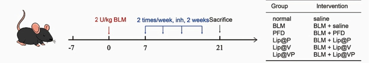
Schematic diagram of the experimental process
Lung function charts showed that the use of dual-drug nanoparticles (Lip@VP) could significantly improve the lung function of fibrosis mice, which was closer to that of normal control mice than other groups of mice.
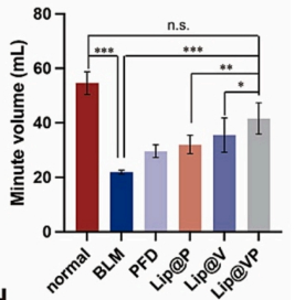
Lung function indicators of mice in each group
The tidal volume, minute ventilation, peak inspiratory flow rate and mid-respiratory flow rate of each group of mice were measured by plethysmography. Compared with the normal group, the other four groups showed a significant decrease, while the Lip@VP group remained almost consistent with the normal group.
The researchers then sectioned and stained the lungs of each group of mice, performing immunofluorescence staining for α-smooth muscle actin (α-SMA) and interstitial protein (krt5). As shown in the figure below:
Lung staining of mice in each group
The image shows that HE staining of the lungs of normal mice reveals intact alveolar structure and a low cell count. However, in mice with fibrosis, the cell count increased significantly, and the alveolar structure became disrupted. After Lip@VP was applied to mice with pulmonary fibrosis, the alveolar structure returned to near-normal. Masson staining revealed significantly darker collagen fibers in fibrotic lung tissue, indicating that the increased number of cells is actually overproliferating fibroblasts. The expression of α-SMA and krt5 was also significantly increased in the lungs of fibrotic mice. Excessive increases in these proteins exacerbate fibrosis, while the Lip@VP group reversed this expression back to normal levels. Quantitative measurements further confirmed that the nanodrug can reduce the overexpression of α-SMA and krt5.
α-SMA and krt5 protein expression bands in each group
The study, through the modeling of pulmonary fibrosis and the reversal of fibrosis with nanomedicines, suggests that pulmonary fibrosis is a process of airway epithelial fluidization and excessive proliferation of fibroblasts. Therefore, inhibiting airway fluidization and fibroblast proliferation is the starting point for treatment. Nanomedicines enter the alveolar surface through intrapulmonary atomization due to their stronger permeability and sustained release ability; VER inhibits airway epithelial fluidization and reduces pathological cell migration, while PFD inhibits excessive fibroblast proliferation. The two work together to reshape the honeycomb cysts and alveolar distortions formed by pulmonary fibrosis, achieving a cure. Overall, the nanoparticle-loaded dual-drug experiment achieved surprising results. By encapsulating the drug in nanoparticles, its penetration and accumulation capacity are enhanced, and by pulmonary atomization, its range of action is increased, finding a new strategy for the clinical treatment of IPF.
Next is an introduction to pulmonary drug delivery.