


Exploring Cardiovascular and Cerebrovascular Disease Research: An Overview of Animal Model Construction and Precision Monitoring Technologies

The middle cerebral artery is the most commonly affected cerebral blood vessel in ischemic stroke among cardiovascular and cerebrovascular diseases, and the middle cerebral artery occlusion (MCAO) model is also one of the most commonly used cardiovascular and cerebrovascular disease models.

In a recent research paper titled "Ion Dyshomeostasis in the Early Hyperacute Phase after a Temporary Large-Vessel Occlusion Stroke," ACS Chemical Neuroscience, a subsidiary of ACS Chemical, a well-known academic journal series published by the American Chemical Society, mice were used to create a MCAO model.

Experimental animals:
11-week-old C57BL/6 male mice.
Modeling method:
Mice were anesthetized with isoflurane gas (induction concentration 3% and maintenance concentration 2%);
After the mice were anesthetized, a midline incision was made in the neck to expose the common carotid artery (CCA);
The external carotid artery (ECA) and internal carotid artery (ICA) were separated, and the ECA was ligated at the distal segment;
A small incision is then made at the bifurcation of the CCA and ICA, and a nylon thread (thread plug) is inserted through the incision into the ICA and advanced to the origin of the middle cerebral artery (MCA).
After the suture was fixed for 30 minutes, the suture was removed to allow blood reperfusion. At the same time, bupivacaine (1 mg/kg/hour of operation time) was dripped into the incision, and then the blood vessels, muscularis, and skin were sutured.
Monitoring methods :
The MouseOx Plus pulse oximeter from Starr Company of the United States was used to monitor the respiratory rate, pulse, and oxygen saturation of the experimental mice and to monitor their vital signs.
Model Validation Methods :
Traditional HE staining was used. Tissues were fixed with 4% paraformaldehyde, washed with 0.1 M phosphate-buffered saline (PBS), and then stained with hematoxylin and eosin (H&E).
Commonly used animal models of cardiovascular disease
In addition to the MCAO model, a typical cerebral ischemia model, many other models are currently widely used in the field of cardiovascular disease research. These models cover a variety of disease types, ranging from hypertension, atherosclerosis to cardiomyopathy.01Hypertension Model
Common hypertension models are divided into two categories: genetic hypertension models and induced hypertension models;
Genetic hypertension models include:
1. Use the hypertensive phenotype to screen for stable inbred models (stroke-prone SIIR hypertensive rats, Dahl/SS salt-sensitive rats, and other selective inbred models)
2. Genetically engineered models (humanized models such as full gene knockout to construct a Mendelian model of hypertension, targeted gene knockout to construct rats with human PDE3A gene mutations, and rats carrying human CACNA1D exon mutations).
Inducible hypertension models are often constructed through surgical or drug induction. For example:
1. Inducing renal vascular hypertension through surgical procedures such as reducing renal artery blood flow, inducing renal parenchymal compression, and subtotal nephrectomy;
2. Long-term subcutaneous injection of Ang II ≥500 ng/(kg·min) can cause an increase in blood pressure in experimental animals after 24 hours, followed by vascular remodeling after 2 weeks, myocardial hypertrophy after 2-4 weeks, and renal damage after 4 weeks.
3. Inducible model using mineralocorticoids, high salt, and nitric oxide synthase inhibitor treatment.
Model validation: Blood pressure can be tested using a non-invasive sphygmomanometer or carotid artery catheterization, and the blood pressure changes of rats can be monitored regularly for model validation.

02Atherosclerosis Mouse Model
Atherosclerosis is the primary pathological process in most cardiovascular diseases. The formation of arterial plaques, arterial stenosis, and arterial occlusion in the late stages is a key indicator of cardiovascular disease risk. In-depth research into the progression of atherosclerosis in humans is significantly limited. Therefore, animal models of atherosclerosis can be used to study pathogenesis and identify potential drug targets for preventing or reversing lesions.
Atherosclerosis model mice are usually gene knockout mice. Common ones include:
2. LDLr knockout mice retain ApoE function and do not produce additional inflammation;
3. ApoE/LDLr double knockout mice can induce hypercholesterolemia model mice that can produce plaques even when fed with normal diet.
Model verification: Ultrasound imaging techniques, such as high-frequency ultrasound, can be used to image the animal's aorta and other blood vessels to observe changes in vascular wall thickness, plaque formation, and hemodynamics.

03 Cardiomyopathy Models in Adults and Mice
Models of dilated cardiomyopathy can be developed through:
2. Biological techniques can also be used to construct cardiac troponin TAK210 mutations to interfere with the cardiac troponin T gene to change myocardial function; some viruses and drugs (B3 coxsackievirus or anthracyclines such as azithromycin) can also induce dilated cardiomyopathy in animals.
Hypertrophic cardiomyopathy disease models can be constructed by encoding different genes and sites such as α-MyHC gene, TnT gene, MyBP-C gene, RLC gene, and a-tropomyosin; disease models can also be obtained by ligating the aortic arch and clamping the left renal artery.
Model Validation: Cardiac function indicators such as left ventricular ejection fraction (LVEF) and left ventricular fractional shortening (LVFS) can be monitored using a small animal echocardiogram. Furthermore, electrocardiograms (ECGs) can be recorded using a polycardiogram to observe abnormalities in cardiac activity, such as arrhythmias.

Yuyan Instruments——Provide comprehensive support for you to build cardiovascular disease animal models
Regardless of the type of cardiovascular disease model being constructed, from hypertension and atherosclerosis to cardiomyopathy and even complex models such as cerebral ischemia, the process is extremely delicate and demanding. Durable and stable surgical instruments and precise and reliable maintenance and testing equipment are indispensable cornerstones of this research and exploration. Shanghai Yuyan Instruments has independently developed a series of high-quality products designed to meet the comprehensive needs of cardiovascular disease model construction.01Surgical Instruments
1.Surgical instrument kit
Yuyan Instruments has developed a variety of surgical instrument kits for researchers. These kits contain a variety of surgical instruments for different application scenarios, eliminating the need for users to select individual instruments, providing a safe and secure environment for animal surgery.Including basic instrument package A4, anatomical separation surgical set D2, microsurgery instrument package F, suture instrument package L, etc.
The Microsurgery Instrument Package F is suitable for myocardial infarction modeling, vascular separation, and other surgical procedures. Other surgical instrument packages are also available to provide strong support for other operations during surgery.

2. Small animal sutures
Yuyan Instruments offers two main categories of surgical sutures for experimental animals: absorbable and non-absorbable. Absorbable sutures are made from PGA and its modified copolymers, offering high strength, flexibility, a suitable hydrolysis cycle, and high biocompatibility. Non-absorbable sutures are made from braided silk, offering excellent flexibility, high strength, stability, and safety. They are suitable for surgical sutures on mice, rats, guinea pigs, rabbits, and larger animals.
Small animal sutures can not only be used for routine surgical sutures, but are also indispensable for vascular ligation and tissue fixation in cardiovascular modeling surgery.

*Suture needles are made of high-purity 302 stainless steel, which has excellent corrosion resistance and biocompatibility;
*High-strength, low-friction suture thread, specially treated to have excellent flexibility, smooth thread body, and smooth suturing process;
*Absorbable PGA sutures do not need to be removed after surgery. Non-absorbable sutures are made of continuous protein fibers from silkworm cocoons and have good biocompatibility. A variety of needle types, curvatures, and sizes are available.
02.Maintain equipment
1. Small animal anesthesia machine
Surgical procedures often require animals to be anesthetized. Inhalation anesthesia has become the mainstream method of animal anesthesia due to its rapid recovery, ease of control, and high surgical success rate. Shanghai Yuyan's inhalation anesthesia system utilizes classic, imported MSS vaporizers that comply with ISO9001 and European CE quality certifications. These vaporizers are individually calibrated at the factory to ensure consistent and stable concentrations.
In cardiovascular and cerebrovascular surgery, the anesthesia machine provides animals with stable anesthetic gas during the operation, keeping the animals in a stable state and contributing to the success of the operation.

*Imported tank, stable output, with temperature compensation function
*Compact design to improve space utilization
*Precise control, stable animal physiological state
2. Body temperature maintenance device
The S-100 body temperature maintenance instrument independently developed and produced by Shanghai Yuyan has a built-in dual-channel heating module that can heat two heating pads at the same time to meet the needs of conducting two experiments at the same time.Equipped with a precise platinum resistance temperature sensor to measure rectal temperature, with high measurement accuracy and good stability.
Under anesthesia, the animal's hypothalamic thermoregulatory activity is suppressed, and it is unable to maintain a stable body temperature as in a normal physiological state. To prevent accidental death due to hypothermia, it is recommended to use a temperature maintenance device while under anesthesia.

03Physiological data monitoring equipment
The NIBP small animal non-invasive blood pressure measurement system is an automated non-invasive small animal blood pressure measurement system independently developed by Yuyan Instruments.The built-in physiological signal acquisition processor is used to collect blood pressure signals and the software analyzes them. It can realize the synchronous recording and result display of single-channel or multi-channel rat tail blood pressure waveforms. All test data can be saved as original files for online or offline analysis.
The equipment is mainly used to measure systolic blood pressure, diastolic blood pressure, mean blood pressure, and heart rate parameters of mice and rats, and can be used to evaluate the modeling effect of hypertension and other models.

2. iWorx Multichannel Physiological Recorder
The iWorx biological signal acquisition and analysis system consists of a data collector, analysis software (LabScribe), and various amplifiers, sensors, lead wires, and electrodes.
Through different combinations of various hardware, parameters such as electrocardiogram, electroencephalogram, electromyography, electrooculography, invasive blood pressure, non-invasive blood pressure, dP/dt, body temperature, muscle tension, respiratory wave, respiratory flow rate, lung function, tissue blood flow, vascular blood flow, nerve potential, oxygen content, carbon dioxide content, blood oxygen saturation, cardiac output, pulse volume, electrical stimulation, evoked potential, etc. can be observed and analyzed.
iWorx's powerful physiological signal acquisition function enables it to verify the modeling effects of various models, such as verifying hypertension models by collecting blood pressure signals, and verifying various heart disease models by collecting electrocardiogram signals.

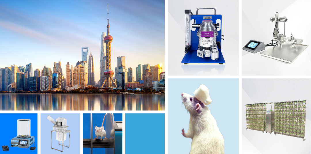
3. MouseOx Plus Pulse Oximeter
Starr Life Sciences' MouseOx Plus pulse oximeter, developed through 20 years of product research and development and technological optimization, is specifically optimized for the high heart rate and high respiratory rate of mice and rats. Using only a single probe, it can accurately obtain pulse oxygen saturation, pulse rate, respiratory rate, pulse amplitude, respiratory amplitude and other parameters through optical signal analysis. By adding an additional temperature probe, the body temperature of animals under anesthesia can also be monitored in real time.
During surgery, the pulse oximeter can collect important physiological signals of animals, helping experimenters understand the current physiological state of the animals. It can also verify some models, such as the ischemia-hypoxia model.

*Accuracy comparable to that of blood gas analyzers;
*Support anesthesia measurement and awake measurement;
* Non-invasive measurement of up to 7 parameters;
* Rich sensor specifications to meet different experimental needs
More product recommendations

For more product inquiries, please contact us!
Self-developed core creates extraordinary strength
R&D personnel account for 40% of the company, and it has a team of scientists in sensors, chip design, core algorithms, etc. There are professional teams to provide support in product implementation and operation, market and academic promotion, comprehensive product solution design and application. The company has service points covering the whole country and strong technical service capabilities. Its customers include Tsinghua University, Peking University, Zhejiang University, Shanghai Jiaotong University, University of Chinese Academy of Sciences, West China Hospital of Sichuan University, Northern Theater Command General Hospital and other first-class domestic and foreign research institutions and hospitals.
- Prev: In addition to intraperitoneal injection, there are so many ways to administer drugs to mice!A complete analysis of drug administration methods for experimental animals
- Next: Small Animal Pulse Oximeters Help Non-Invasive and Accurate Blood Oxygen Measurement in Animals with Lung Injury!(Detailed experimental methods are included)