


Peira | Small Animal Tumor Measuring Instrument - Say Goodbye to Manual Measurement!Black technology for measuring surface tumor size
Date:2024-04-09
Author:Yuyan Instrument
Cancer, a major killer threatening human health, has long been a focus of medical research on its pathogenesis and treatment. With advances in science and technology, the research, development, and application of anticancer drugs have achieved remarkable results in recent years.
From the initial chemotherapy drugs to today's targeted therapies, immunotherapy, etc., anti-cancer drugs have been continuously upgraded, bringing more treatment options and hope to cancer patients.
However, the development and application of anticancer drugs still face numerous challenges, such as side effects and drug resistance, which need to be addressed urgently. Therefore, in-depth research on the therapeutic mechanisms of anticancer drugs, optimizing treatment options, and improving treatment efficacy are of great significance for prolonging patient survival and improving quality of life.
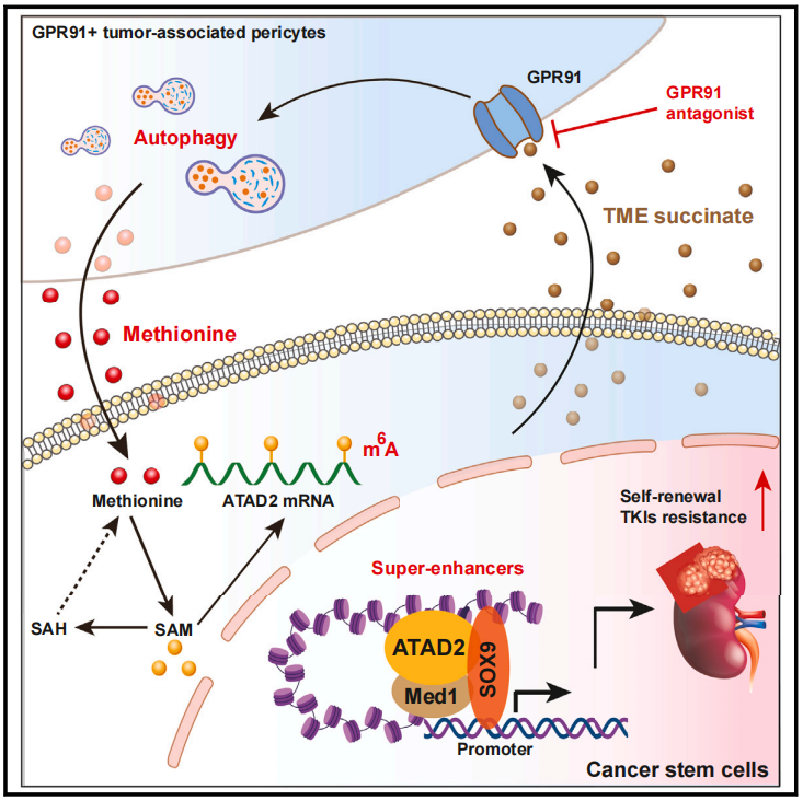
On February 19, 2024, Professor Li Qingquan's team from Fudan University published a research paper titled "Methionine secreted by tumor-associated pericytes supports cancer stem cells in clear cell renal carcinoma" in the journal Cell Metabolism. This research is of great significance for the development of drugs to treat ccRCC (clear cell renal cell carcinoma).
Here we identify a subpopulation of vascular pericytes, defined by expression of platelet-derived growth factor receptor β (PDGFR-β) and G protein-coupled receptor 91 (GPR91), that promotes tumorigenesis and resistance to tyrosine kinase inhibitors (TKIs) by serving as a major source of methionine for cancer stem cells (CSCs) in clear cell renal cell carcinoma (ccRCC). Tumor cell-derived succinate binds to GPR91 on pericytes, activating autophagy to generate methionine.
The article primarily elucidates that CSCs utilize methionine to generate stable N6-methyladenosine in ATPase-family-AAA-domain-containing 2 (ATAD2) mRNA. The resulting ATAD2 protein complex assembles with SRY-box transcription factor 9 to form a super-enhancer, thereby instructing its target genes to be prominently characterized in CSCs. A specific GRP91 antagonist targets PDGFR-β+GPR91+ pericytes, reducing intratumoral methionine levels, eliminating CSCs, and enhancing TKI sensitivity. These results reveal a mechanism by which PDGFR-β+GPR91+ pericytes provide a supportive niche for CSCs and may be used to develop therapeutic targets for ccRCC.
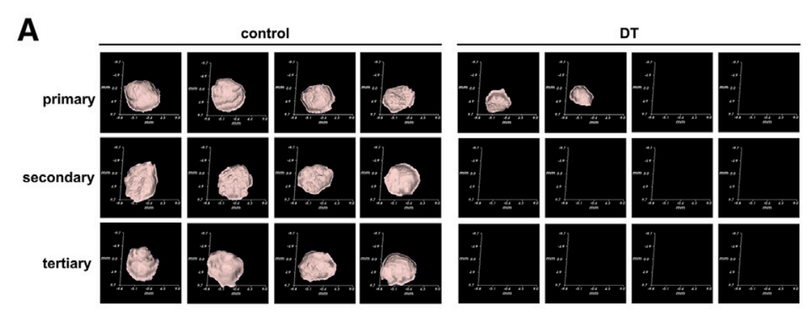
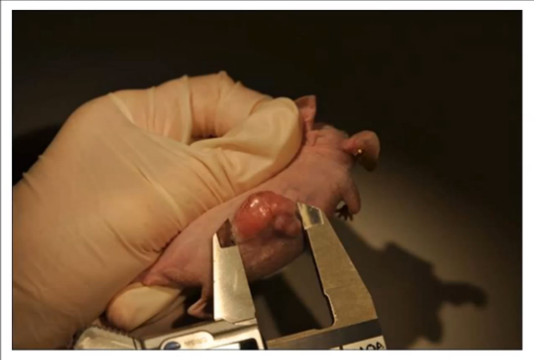
In the study of the mechanism of this pericyte subset, in order to study the significance of PDGFR-b+GPR91+ pericytes to renal cancer stem cells, the author team used the Peira TM900 tumor measuring instrument to measure the tumor frequency (n=4) after subcutaneous injection of NOD/SCID mice with NOD or dt-treated Ksp1.3-tva;PGDTR mice with tumor cells (105). It was found that the continuous transplantation ability of tumors in treated mice was significantly reduced. The Peira TM900 tumor measuring instrument can directly present the three-dimensional effect of the tumor and measure related parameters, which greatly reduces the workload and measurement error of manual measurement.
Tumor size measurement image derived from Peira TM900
Caliper measurement method
This method is applicable to postmortem measurement of solid tumors located on the surface of animals and solid tumors located in the body after animal euthanasia. The tumor volume calculated as an ellipsoid, as mentioned in a 1989 paper by Mary M. Tomayko and C. Patrick Reynolds of the University of California, Los Angeles School of Medicine, is often used:
V=π/6×L (long diameter)×W (short diameter)×H (height)
Before using this method, the tumor's long and short axes should be determined to ensure they are perpendicular during measurement. For subcutaneous tumor measurement, use calipers to measure length (L, the long dimension), width (W, the short dimension, perpendicular to the length plane and parallel to the animal's body), and height (H, the distance between the upper border of the tumor and the animal's body).
This method has many problems. First, the length and width are difficult to judge. Second, the height of the tumor is not easy to measure because it grows out of the skin on the other side. In addition, different experimenters have different ways of judging the length and using vernier calipers, resulting in large differences in measurements.
Imaging equipment imaging method
Various animal-specific imaging devices enable real-time, continuous, non-invasive, and in situ observation and measurement of tumors. Currently used in animal model research are ultrasound, micro-CT (micro CT), PET (micro PT), in vivo small animal imaging, and micro-magnetic resonance imaging (micro MRI). All of these devices can image and measure tumors, but they are expensive and often require dedicated rooms and personnel, resulting in high maintenance costs.
Tumor measuring instrument
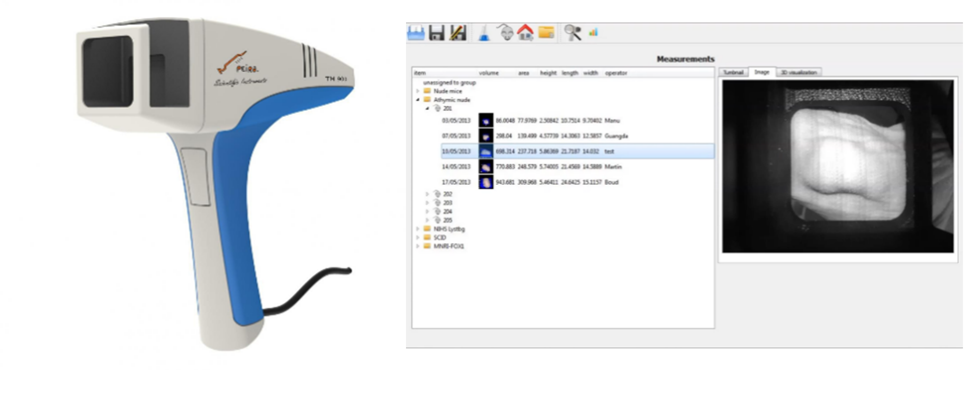
The Peira TM900 Tumor Meter, a cutting-edge technology for tumor measurement, offers researchers a new and superior option for tumor size measurement. The Peira TM900 Tumor Meter, powered by advanced stereoscopic imaging technology, eliminates the need for calipers or other imaging equipment to capture and generate 3D tumor images. When structured light from the tumor is projected onto the scanner's acquisition port, the system automatically calculates the tumor's 3D image and volume based on the deformation coefficients of the structured light, eliminating the need for manual measurement or calculation.
The TM900 Tumor Meter is an advanced device designed specifically for scientific research, used to precisely measure tumor size and morphology. Combining modern imaging technology with computer image processing techniques, the instrument provides accurate and reliable tumor size data, providing strong support for tumor research.
Designed to address the problems of cumbersome operations and highly subjective data in the tumor measurement process, the instrument has a handheld imaging device. Experimenters only need to pick up the measuring probe, aim it at the tumor site, and measure it to quickly create a three-dimensional image of the tumor and present it on the computer.
Based on advanced stereo imaging technology, the system captures 3D tumor images. When the tumor's structured light is projected onto the scanner's acquisition port, the system automatically calculates the tumor's 3D image and volume based on the deformation coefficient of the structured light. Software then creates a 3D image of the tumor and calculates its size, volume, and other data.
2. Handheld design, simple and fast
The probe is handheld and equipped with a computer. The handheld design allows for measurement at any angle, with flexible measurement methods and full coverage of the measurement process without visible light interference. The measurement can be started by pressing the trigger button, and there will be sound and light to indicate the end of the measurement. It is convenient and quick to use.
3. Multi-size adaptation to meet different size requirements
Tumors of any size can be easily measured. The instrument is equipped with a variety of probe covers, which are suitable for measuring tumors of different sizes, avoiding excessively long reflection cycles of structured light, while ensuring full coverage of the measurement process and avoiding the influence of visible light on the measurement.
4. Professional software analysis, highly visualized
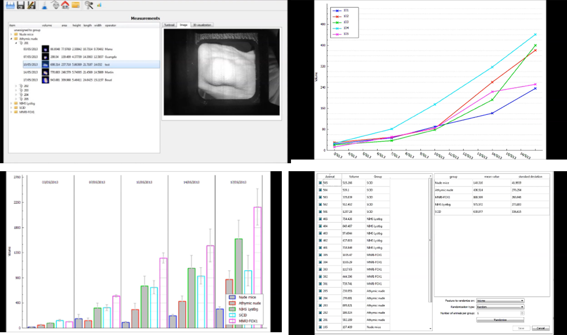
The integrated software package provides researchers with automated tumor data collection, storing volume, area, height, real-world images, and 3D modeling data for long-term follow-up observation and multi-parameter comparison. The software displays tumor trends and comparisons for each animal or group, tracking the entire experimental process without manual organization or calculations.
Volume, area, height, real image, and 3D modeling image can be saved for long-term tracking observation and to meet multi-parameter comparison requirements.
The integrated software package allows researchers to automatically collect tumor data, display tumor trends for each mouse or group, and track the progress of the entire experiment without having to organize it themselves.
The data can be exported in various formats, such as csv, pdf, bmp, png, etc., which can meet the needs of later data processing.
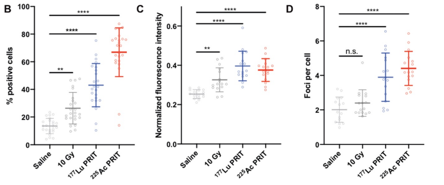
DNA double-strand break 89Zr-PET imaging for early monitoring of the efficacy of α- and β-particle radioimmunotherapy in pancreatic ductal adenocarcinoma
Research topics
This study investigated the effects of α- and β-particle radioimmunotherapy (PRIT) in a mouse model of pancreatic ductal adenocarcinoma (PDAC) and assessed this using PET imaging. The DNA damage marker γH2AX was significantly increased after PRIT treatment, but PET imaging did not reveal differences in the radiobiology of α- and β-PRIT tumors. This suggests that DNA damage is not the sole radiobiological mechanism and that bystander effects should be considered.
To ensure consistency in the grouping of tumor models, the author team used the Peira TM900 tumor measuring instrument to measure the tumor size of each model mouse and randomly grouped the mice based on the measurement results of the software, greatly improving the statistical credibility of the experiment.
——Poty, Sophie et al. “89Zr-PET imaging of DNA double-strand breaks for the early monitoring of response following α- and β-particle radioimmunotherapy in a mouse model of pancreatic ductal adenocarcinoma.” Theranostics vol. 10,13 5802-5814. 27 Apr. 2020, doi:10.7150/thno.44772
Case 2
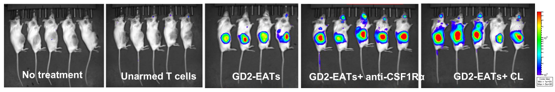
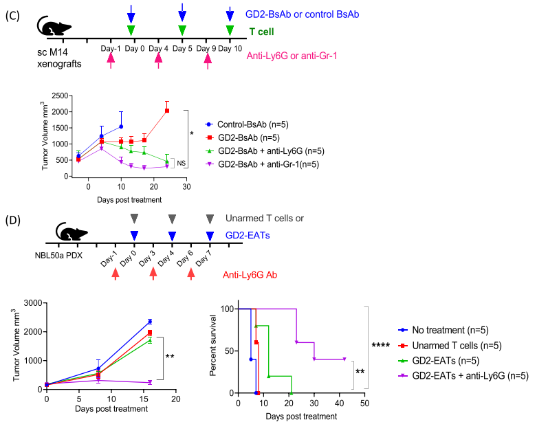
Modulating tumor-infiltrating myeloid cells to enhance bispecific antibody-driven T cell infiltration and antitumor responses
Research topics
This study investigated the role of GD2-BsAb and granulocyte-depleting antibodies in cancer therapy. The study found that GD2-BsAb and granulocyte-depleting antibodies enhanced BsAb-guided T cell infiltration and improved anti-tumor efficacy. Furthermore, GD2-BsAb and granulocyte-depleting antibodies reduced the number of immunosuppressive cells in the tumor microenvironment, such as M2 macrophages and tumor-associated macrophages. These results suggest that GD2-BsAb and granulocyte-depleting antibodies have potential application in cancer immunotherapy.
To monitor tumor growth in mice inoculated with tumor cells, the author team used the Peira TM900 tumor measuring instrument to measure the size of the mice's tumors and monitored the tumor growth process. Through measurement, mice that did not meet the conditions were excluded and the tumor growth process was quantitatively analyzed, greatly improving the efficiency of the experiment.
——Park, Jeong A et al. “Modulating tumor infiltrating myeloid cells to enhance bispecific antibody-driven T cell infiltration and anti-tumor response.” Journal of hematology & oncology vol. 14,1 142. 8 Sep. 2021, doi:10.1186/s13045-021-01156-5
As the general agent of Peira in China, Shanghai Yuyan Scientific Instrument Co., Ltd. works together to provide domestic users with advanced and reliable scientific research instrument platforms and provide scientific researchers with internationally leading oncology research instrument solutions.
2. Adams, Elizabeth J et al. “FOXA1 mutations alter pioneering activity, differentiation and prostate cancer phenotypes.” Nature vol. 571,7765 (2019): 408-412. doi:10.1038/s41586-019-1318-9
3. Henry, Kelly E et al. “A PET Imaging Strategy for Interrogating Target Engagement and Oncogene Status in Pancreatic Cancer.” Clinical cancer research : an official journal of the American Association for Cancer Research vol. 25,1 (2019): 166-176. doi:10.1158/1078-0432.CCR-18-1485
4. Park, Jeong A et al. "Modulating tumor infiltrating myeloid cells to enhance bispecific antibody-driven T cell infiltration and anti-tumor response." Journal of hematology & oncology vol. 14,1 142. 8 Sep. 2021, doi:10.1186/s13045-021-01156-5
5. Park, Jeong A, and Nai-Kong V Cheung. "GD2 or HER2 targeting T cell engaging bispecific antibodies to treat osteosarcoma." Journal of hematology & oncology vol. 13,1 172. 10 Dec. 2020, doi:10.1186/s13045-020-01012-y
6. Mao, Ninghui et al. “Defining the therapeutic selective dependencies for distinct subtypes of PI3K pathway-altered prostate cancers.” Nature communications vol. 12,1 5053. 20 Aug. 2021, doi:10.1038/s41467-021-25341-9
7. Wang, Jin-Yan et al. "Immunotherapy combining tumor and endothelium cell lysis with immune enforcement by recombinant MIP-3α Newcastle disease virus in a vessel-targeting liposome enhances antitumor immunity." Journal for immunotherapy of cancer vol. 10,3 (2022): e003950. doi:10.1136/jitc-2021-003950
8. Zhang, Zeda et al. “Tumor Microenvironment-Derived NRG1 Promotes Antiandrogen Resistance in Prostate Cancer.” Cancer cell vol. 38,2 (2020): 279-296.e9. doi:10.1016/j.ccell.2020.06.005
9. Wang, Cheng-Kai et al. “MEX3A Mediates p53 Degradation to Suppress Ferroptosis and Facilitate Ovarian Cancer Tumorigenesis.” Cancer research vol. 83,2 (2023): 251-263. doi:10.1158/0008-5472.CAN-22-1159
10. Szymańska, Ewelina et al. “Synthetic lethality between VPS4A and VPS4B triggers an inflammatory response in colorectal cancer.” EMBO molecular medicine vol. 12,2 (2020): e10812. doi:10.15252/emmm.201910812
From the initial chemotherapy drugs to today's targeted therapies, immunotherapy, etc., anti-cancer drugs have been continuously upgraded, bringing more treatment options and hope to cancer patients.
However, the development and application of anticancer drugs still face numerous challenges, such as side effects and drug resistance, which need to be addressed urgently. Therefore, in-depth research on the therapeutic mechanisms of anticancer drugs, optimizing treatment options, and improving treatment efficacy are of great significance for prolonging patient survival and improving quality of life.

On February 19, 2024, Professor Li Qingquan's team from Fudan University published a research paper titled "Methionine secreted by tumor-associated pericytes supports cancer stem cells in clear cell renal carcinoma" in the journal Cell Metabolism. This research is of great significance for the development of drugs to treat ccRCC (clear cell renal cell carcinoma).
Here we identify a subpopulation of vascular pericytes, defined by expression of platelet-derived growth factor receptor β (PDGFR-β) and G protein-coupled receptor 91 (GPR91), that promotes tumorigenesis and resistance to tyrosine kinase inhibitors (TKIs) by serving as a major source of methionine for cancer stem cells (CSCs) in clear cell renal cell carcinoma (ccRCC). Tumor cell-derived succinate binds to GPR91 on pericytes, activating autophagy to generate methionine.
The article primarily elucidates that CSCs utilize methionine to generate stable N6-methyladenosine in ATPase-family-AAA-domain-containing 2 (ATAD2) mRNA. The resulting ATAD2 protein complex assembles with SRY-box transcription factor 9 to form a super-enhancer, thereby instructing its target genes to be prominently characterized in CSCs. A specific GRP91 antagonist targets PDGFR-β+GPR91+ pericytes, reducing intratumoral methionine levels, eliminating CSCs, and enhancing TKI sensitivity. These results reveal a mechanism by which PDGFR-β+GPR91+ pericytes provide a supportive niche for CSCs and may be used to develop therapeutic targets for ccRCC.
In the study of the mechanism of this pericyte subset, in order to study the significance of PDGFR-b+GPR91+ pericytes to renal cancer stem cells, the author team used the Peira TM900 tumor measuring instrument to measure the tumor frequency (n=4) after subcutaneous injection of NOD/SCID mice with NOD or dt-treated Ksp1.3-tva;PGDTR mice with tumor cells (105). It was found that the continuous transplantation ability of tumors in treated mice was significantly reduced. The Peira TM900 tumor measuring instrument can directly present the three-dimensional effect of the tumor and measure related parameters, which greatly reduces the workload and measurement error of manual measurement.
Tumor size measurement image derived from Peira TM900
Tumor size measurement methods
In tumor model animals, tumors often grow into spherical, elliptical, long strips, or even irregular shapes, which makes it difficult to measure tumor volume. Therefore, the description of tumor size in living animals is often an estimate. There are currently many methods to measure tumor size in preclinical studies.Caliper measurement method
This method is applicable to postmortem measurement of solid tumors located on the surface of animals and solid tumors located in the body after animal euthanasia. The tumor volume calculated as an ellipsoid, as mentioned in a 1989 paper by Mary M. Tomayko and C. Patrick Reynolds of the University of California, Los Angeles School of Medicine, is often used:
V=π/6×L (long diameter)×W (short diameter)×H (height)
Before using this method, the tumor's long and short axes should be determined to ensure they are perpendicular during measurement. For subcutaneous tumor measurement, use calipers to measure length (L, the long dimension), width (W, the short dimension, perpendicular to the length plane and parallel to the animal's body), and height (H, the distance between the upper border of the tumor and the animal's body).
This method has many problems. First, the length and width are difficult to judge. Second, the height of the tumor is not easy to measure because it grows out of the skin on the other side. In addition, different experimenters have different ways of judging the length and using vernier calipers, resulting in large differences in measurements.
Imaging equipment imaging method
Various animal-specific imaging devices enable real-time, continuous, non-invasive, and in situ observation and measurement of tumors. Currently used in animal model research are ultrasound, micro-CT (micro CT), PET (micro PT), in vivo small animal imaging, and micro-magnetic resonance imaging (micro MRI). All of these devices can image and measure tumors, but they are expensive and often require dedicated rooms and personnel, resulting in high maintenance costs.
Tumor measuring instrument
The Peira TM900 Tumor Meter, a cutting-edge technology for tumor measurement, offers researchers a new and superior option for tumor size measurement. The Peira TM900 Tumor Meter, powered by advanced stereoscopic imaging technology, eliminates the need for calipers or other imaging equipment to capture and generate 3D tumor images. When structured light from the tumor is projected onto the scanner's acquisition port, the system automatically calculates the tumor's 3D image and volume based on the deformation coefficients of the structured light, eliminating the need for manual measurement or calculation.
Peira TM900 tumor measurement instrument brings you an excellent new experience in tumor measurement
Instrument IntroductionThe TM900 Tumor Meter is an advanced device designed specifically for scientific research, used to precisely measure tumor size and morphology. Combining modern imaging technology with computer image processing techniques, the instrument provides accurate and reliable tumor size data, providing strong support for tumor research.
Designed to address the problems of cumbersome operations and highly subjective data in the tumor measurement process, the instrument has a handheld imaging device. Experimenters only need to pick up the measuring probe, aim it at the tumor site, and measure it to quickly create a three-dimensional image of the tumor and present it on the computer.
Main Features
1. 3D scanning imaging, automatic measurement data is reliableBased on advanced stereo imaging technology, the system captures 3D tumor images. When the tumor's structured light is projected onto the scanner's acquisition port, the system automatically calculates the tumor's 3D image and volume based on the deformation coefficient of the structured light. Software then creates a 3D image of the tumor and calculates its size, volume, and other data.
2. Handheld design, simple and fast
The probe is handheld and equipped with a computer. The handheld design allows for measurement at any angle, with flexible measurement methods and full coverage of the measurement process without visible light interference. The measurement can be started by pressing the trigger button, and there will be sound and light to indicate the end of the measurement. It is convenient and quick to use.
3. Multi-size adaptation to meet different size requirements
Tumors of any size can be easily measured. The instrument is equipped with a variety of probe covers, which are suitable for measuring tumors of different sizes, avoiding excessively long reflection cycles of structured light, while ensuring full coverage of the measurement process and avoiding the influence of visible light on the measurement.
4. Professional software analysis, highly visualized
The integrated software package provides researchers with automated tumor data collection, storing volume, area, height, real-world images, and 3D modeling data for long-term follow-up observation and multi-parameter comparison. The software displays tumor trends and comparisons for each animal or group, tracking the entire experimental process without manual organization or calculations.
Supporting software
The instrument is equipped with a computer with built-in management and measurement software. The data management software can compile animal names, groups, cages, and individuals in four levels.Volume, area, height, real image, and 3D modeling image can be saved for long-term tracking observation and to meet multi-parameter comparison requirements.
The integrated software package allows researchers to automatically collect tumor data, display tumor trends for each mouse or group, and track the progress of the entire experiment without having to organize it themselves.
The data can be exported in various formats, such as csv, pdf, bmp, png, etc., which can meet the needs of later data processing.
More literature cases
Case 1DNA double-strand break 89Zr-PET imaging for early monitoring of the efficacy of α- and β-particle radioimmunotherapy in pancreatic ductal adenocarcinoma
Research topics
This study investigated the effects of α- and β-particle radioimmunotherapy (PRIT) in a mouse model of pancreatic ductal adenocarcinoma (PDAC) and assessed this using PET imaging. The DNA damage marker γH2AX was significantly increased after PRIT treatment, but PET imaging did not reveal differences in the radiobiology of α- and β-PRIT tumors. This suggests that DNA damage is not the sole radiobiological mechanism and that bystander effects should be considered.
To ensure consistency in the grouping of tumor models, the author team used the Peira TM900 tumor measuring instrument to measure the tumor size of each model mouse and randomly grouped the mice based on the measurement results of the software, greatly improving the statistical credibility of the experiment.
——Poty, Sophie et al. “89Zr-PET imaging of DNA double-strand breaks for the early monitoring of response following α- and β-particle radioimmunotherapy in a mouse model of pancreatic ductal adenocarcinoma.” Theranostics vol. 10,13 5802-5814. 27 Apr. 2020, doi:10.7150/thno.44772
Case 2
Modulating tumor-infiltrating myeloid cells to enhance bispecific antibody-driven T cell infiltration and antitumor responses

Research topics
This study investigated the role of GD2-BsAb and granulocyte-depleting antibodies in cancer therapy. The study found that GD2-BsAb and granulocyte-depleting antibodies enhanced BsAb-guided T cell infiltration and improved anti-tumor efficacy. Furthermore, GD2-BsAb and granulocyte-depleting antibodies reduced the number of immunosuppressive cells in the tumor microenvironment, such as M2 macrophages and tumor-associated macrophages. These results suggest that GD2-BsAb and granulocyte-depleting antibodies have potential application in cancer immunotherapy.
To monitor tumor growth in mice inoculated with tumor cells, the author team used the Peira TM900 tumor measuring instrument to measure the size of the mice's tumors and monitored the tumor growth process. Through measurement, mice that did not meet the conditions were excluded and the tumor growth process was quantitatively analyzed, greatly improving the efficiency of the experiment.
——Park, Jeong A et al. “Modulating tumor infiltrating myeloid cells to enhance bispecific antibody-driven T cell infiltration and anti-tumor response.” Journal of hematology & oncology vol. 14,1 142. 8 Sep. 2021, doi:10.1186/s13045-021-01156-5
About Peira

Peira is a brand of Komax, a Belgian company, dedicated to the development of equipment for preclinical drug development. It has professional researchers in the fields of in vivo, ex vivo and in vitro experimental devices and imaging automation, and has extensive experience in equipment research in fields such as neuroscience, oncology and toxicology.
As the general agent of Peira in China, Shanghai Yuyan Scientific Instrument Co., Ltd. works together to provide domestic users with advanced and reliable scientific research instrument platforms and provide scientific researchers with internationally leading oncology research instrument solutions.
Further Reading
1. Hadi, Marym Mohammad et al. “Investigating the performance of a novel pH and cathepsin B sensitive, stimulus-responsive nanoparticle for optimized sonodynamic therapy in prostate cancer.” Journal of controlled release : official journal of the Controlled Release Society vol. 329 (2021): 76-86. doi:10.1016/j.jconrel.2020.11.0402. Adams, Elizabeth J et al. “FOXA1 mutations alter pioneering activity, differentiation and prostate cancer phenotypes.” Nature vol. 571,7765 (2019): 408-412. doi:10.1038/s41586-019-1318-9
3. Henry, Kelly E et al. “A PET Imaging Strategy for Interrogating Target Engagement and Oncogene Status in Pancreatic Cancer.” Clinical cancer research : an official journal of the American Association for Cancer Research vol. 25,1 (2019): 166-176. doi:10.1158/1078-0432.CCR-18-1485
4. Park, Jeong A et al. "Modulating tumor infiltrating myeloid cells to enhance bispecific antibody-driven T cell infiltration and anti-tumor response." Journal of hematology & oncology vol. 14,1 142. 8 Sep. 2021, doi:10.1186/s13045-021-01156-5
5. Park, Jeong A, and Nai-Kong V Cheung. "GD2 or HER2 targeting T cell engaging bispecific antibodies to treat osteosarcoma." Journal of hematology & oncology vol. 13,1 172. 10 Dec. 2020, doi:10.1186/s13045-020-01012-y
6. Mao, Ninghui et al. “Defining the therapeutic selective dependencies for distinct subtypes of PI3K pathway-altered prostate cancers.” Nature communications vol. 12,1 5053. 20 Aug. 2021, doi:10.1038/s41467-021-25341-9
7. Wang, Jin-Yan et al. "Immunotherapy combining tumor and endothelium cell lysis with immune enforcement by recombinant MIP-3α Newcastle disease virus in a vessel-targeting liposome enhances antitumor immunity." Journal for immunotherapy of cancer vol. 10,3 (2022): e003950. doi:10.1136/jitc-2021-003950
8. Zhang, Zeda et al. “Tumor Microenvironment-Derived NRG1 Promotes Antiandrogen Resistance in Prostate Cancer.” Cancer cell vol. 38,2 (2020): 279-296.e9. doi:10.1016/j.ccell.2020.06.005
9. Wang, Cheng-Kai et al. “MEX3A Mediates p53 Degradation to Suppress Ferroptosis and Facilitate Ovarian Cancer Tumorigenesis.” Cancer research vol. 83,2 (2023): 251-263. doi:10.1158/0008-5472.CAN-22-1159
10. Szymańska, Ewelina et al. “Synthetic lethality between VPS4A and VPS4B triggers an inflammatory response in colorectal cancer.” EMBO molecular medicine vol. 12,2 (2020): e10812. doi:10.15252/emmm.201910812
- Prev: Ugo Basile's startle reflex system | An important experimental tool for studying the mechanisms of mental illness, antipsychotic drugs, and sensorimotor gating function
- Next: Pinnacle | A one-stop epilepsy monitoring and analysis system for small animals, enabling high-throughput screening models for anti-epileptic drugs to achieve new results