


Manipulating brain activity: Optogenetics brings neuronal control from science fiction to reality
Classic movie plot: manipulating the brain
If you have seen the movie "Lucy", you must be impressed by this scene - Lucy, a young woman who was forced to help deliver goods, encountered a group of vicious gang leaders. Mr. Zhang had a mysterious drug sewn into her abdomen to smuggle the drug into different countries. While staying in Taipei, Lucy was beaten so badly that the drug broke and entered her blood.Unexpectedly, something incredible happened. The drug stimulated the potential of Lucy's brain, and about 90% of the neurons in her brain were awakened one after another. With the rapid evolution of her body, Lucy mastered more and more so-called super powers.
Science fiction about various ways to control brain activity has been popular for decades. In fact, some of these methods, such as drugs, electrical stimulation, and magnetic stimulation, have been widely used in human clinical practice.

Can light control the brain?
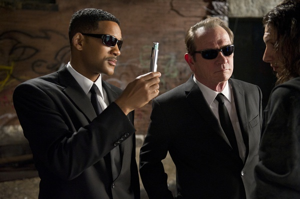
In previous science fiction films, human brain control was primarily achieved through various means, including chemicals, thoughts, and electrical stimulation. However, in the 1997 film "Men in Black," we see a scene where an agent, after handling an alien infestation, pulls out a glow stick and tells the onlookers to "look here." A flash of light then erases the onlookers' short-term memories, obliterating their memories of ever having seen a strange alien creature. It's easy to see that over two decades ago, humans were exploring and imagining how light could control the brain, and at the time, it was just a science fiction theme for movies.
The emergence of optogenetics
In fact, our scientists have been exploring the relationship between light and the brain since the beginning of the 21st century.In 2002, Professor Gero Miesenböck of the Memorial Sloan Kettering Cancer Center took the lead in trying this bold idea. He expressed light-sensitive proteins from invertebrates in rat cells and saw neurons responding to light stimulation in a culture dish.
In 2005, Karl Deisseroth's laboratory at Stanford University expressed light-sensitive proteins in nerve cells, which responded to light stimulation of different wavelengths to regulate neural function, announcing that humans officially had the tools to precisely control the brain.
In 2006, Karl Deisseroth first proposed optogenetics and, together with 26-year-old Edward Boyden, pioneered the era of optogenetics.
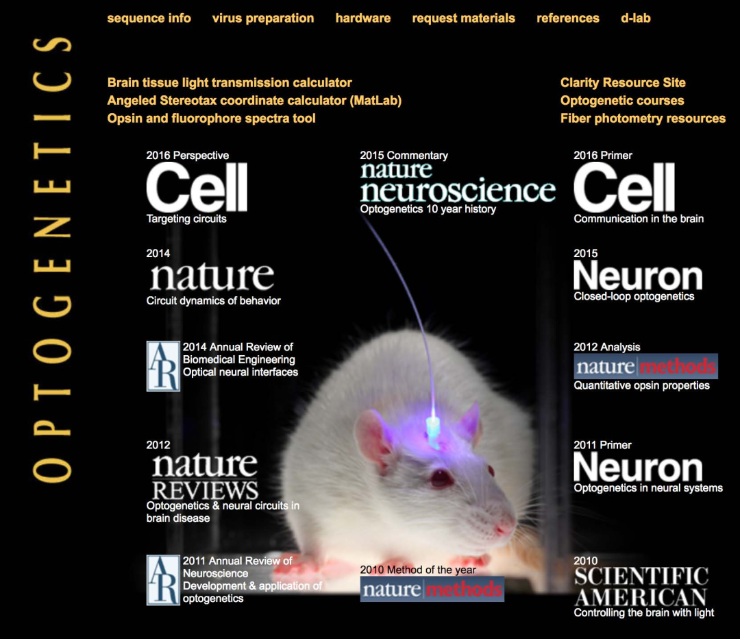
Nature Methods magazine selected Optogenetics as its Research Method of the Year for 2010, and launched a commemorative special issue on its tenth anniversary, commenting on the top ten technologies that have had the greatest impact on biological research in the past decade, including optogenetics.
In 2015, Nature Neuroscience magazine published an article commemorating the tenth anniversary of optogenetics, stating that "optogenetics has opened the door to long-awaited experiments." Some scientists even predicted that "optogenetics is destined to win a Nobel Prize."
The emergence of optogenetics
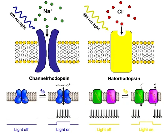
In classic biological experiments, controlling nerve cells requires some relatively rough means, such as electrical stimulation, brain damage or chemical intervention. The experimental results are often unsatisfactory, but optogenetics can solve this problem by controlling single neurons.Optogenetics is a cell biology research method that combines genetics (recombinant DNA technology) with optics. It usually uses viral vectors or transgenic technology to express light receptor (light-sensitive protein) ion channels in receptor cells, and uses light of specific wavelengths to directionally activate or inhibit the cell's discharge activity.
Experimental Procedure
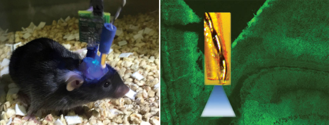
Step 1: Express the light-sensitive protein gene in the target cells: Transfect the virus carrying the light-sensitive protein into the target cells, and express the light-sensitive protein on the cell membrane through the promoter.Step 2: Implant a fiber optic catheter or optogenetic electrode: Implant a fiber optic catheter at the site of virus injection and input light stimulation of a specific wavelength through the fiber optic catheter to activate or inhibit the photosensitive protein.
Step 3: Collect the physiological effects of light stimulation: After light stimulation, cells, tissues or organisms will show corresponding physiological or behavioral changes. The changes in different dimensions caused by optogenetics can be recorded through EEG and EMG acquisition, brain neurotransmitter detection or behavioral video acquisition.
In early optogenetic experiments, light stimulation was usually applied to large areas of the animal's brain, which was far more intense than the experiment required. With the improvement of optogenetic tools and the more precise location of the experimental subject's brain area, more precise light stimulation through optical fibers is becoming the key to optogenetic technology.
Pinnacle optogenetic system in the United States
It should be noted that the optogenetic system is only a research tool, a "switch" that uses light to control brain neural activity, and it does not have a detection function itself.When scientists conduct animal brain research, in order to detect the effects of light stimulation, they often need to use modules such as EEG and EMG acquisition, brain neurotransmitter detection, and behavioral video recording for multi-dimensional analysis.
The Pinnacle optogenetics system in the United States integrates professional EEG and EMG acquisition, neurotransmitter detection, and behavioral video acquisition modules into a highly compatible one-stop system based on the optogenetic stimulation module. It can support researchers in the entire experimental process from implanting fiber optic probes, implementing specific wavelength light stimulation, to collecting physiological and behavioral changes of animals before and after stimulation.

Similar devices on the market have single functions, while the Pinnacle optogenetics system in the United States can perform different tests due to its high scalability.
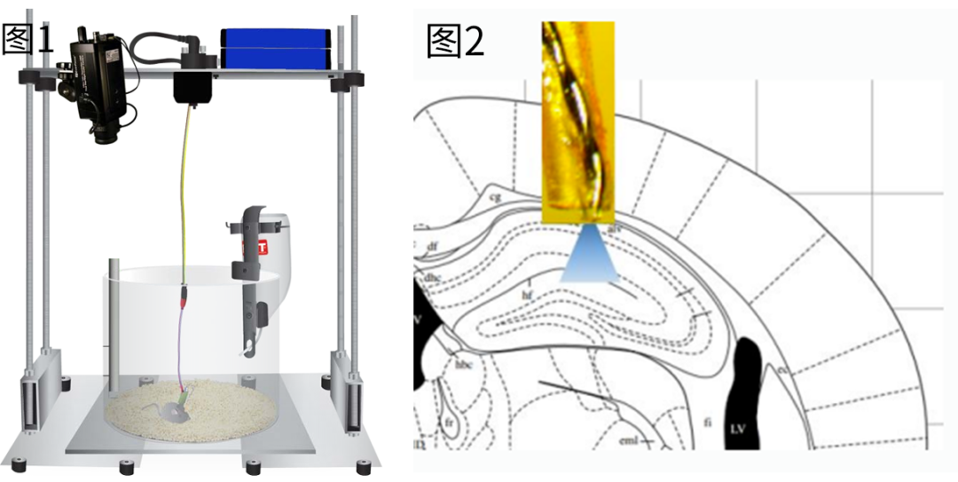
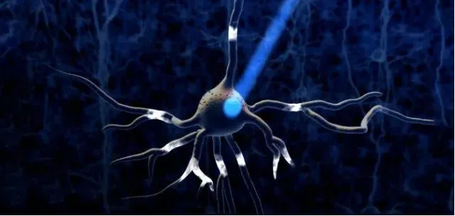
(Figure 1) Behavioral video acquisition + neurotransmitter detection + EEG and EMG acquisition + light stimulation system composition (Figure 2) Light stimulation of neuronal activity
System Features
1. It can be freely used with EEG and myoelectric signal acquisition, neurotransmitter detection, and behavioral video acquisition systems, with high configuration flexibility2. Optical stimulation uses an electrical converter to drive the optical fiber probe, without the need for an additional optical converter
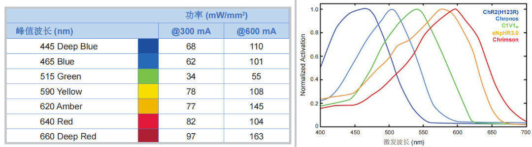
3. LED probes with multiple wavelengths are optional. If you need to change to a different wavelength of light stimulation, you only need to replace the probe. It has a wide range of applications and low cost.
4. High time control accuracy, up to millisecond or sub-millisecond level
5. Strong spatial specificity of light stimulation and specificity for channel proteins
6. LED light source acts directly, and the light stimulation and thermal effect cause little damage to animals
Optogenetics Research System Application Solutions
Optogenetics system combined with EEG and EMG acquisition systemThe innacle fiber optic probe's cannula and prefabricated head cap are fixed to the animal's skull with dental cement, and EEG electrodes are implanted in the brain's corresponding location for light stimulation or in the cortex to collect the animal's EEG signals.
The system can measure neuronal discharge activity before, during, and after light stimulation, accurately recording the activation or inhibition of neurons by light stimulation
Optogenetics system paired with biosensors
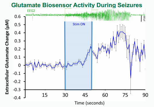
Optogenetics combined with biosensors can detect changes in the concentration of neurotransmitters in the brain while giving light stimulation, which can better study the relationship between brain neuronal network function and behavior. The figure below shows the experimental results of combining optogenetics technology with EEG acquisition system and biosensors.
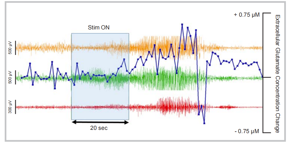
Two-channel electrocortical electroencephalogram (EEG1 and EEG2) recording electrodes were surgically implanted in the brain region of transgenic mice expressing rhodopsin-2, an LED light probe and deep electrodes were implanted in the right hippocampus, and a glutamate biosensor was implanted in the right frontal cortex.
One week after the surgery, a 20-second blue (445nm) light stimulation was applied. During and after the stimulation, the electrodes of the three channels recorded obvious epileptic seizure activity. This is because the corresponding neurons were activated by the optogenetic method. The biosensor detected the release of glutamate in the right frontal cortex at a sub-second level. The real-time sub-second data changes can be perfectly synchronized with the precise timing of the optogenetic stimulation. The data showed that the concentration change of glutamate is closely related to the epileptic seizure. The glutamate concentration drops sharply after the epileptic seizure event.
Optogenetics combined with FSCV system
Pinnacle's integrated optogenetics/fast scan cyclic voltammetry (FSCV) system provides a simple and effective solution for combining optogenetics with FSCV neurotransmitter detection in mice and rats.Researchers can seamlessly integrate optogenetics with electrochemical recordings to detect neurotransmitter release in animal brains, providing a powerful tool for electrochemical detection of neurotransmitters before and after light stimulation in optogenetic experiments.
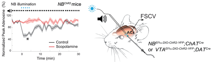
Figure: Optogenetic stimulation experiments were performed on blank control mice and scopolamine-treated mice, and the changes in adenosine concentration in the brains of the two groups of mice were recorded before, during, and after NB light stimulation.
More application cases
Case 1
A virus encoding channelrhodopsin-2 was injected into the deep and intermediate layers of the superior colliculus of rats, and a head-mounted LED fiber optic probe was implanted in the same location. Pilocarpine was used to induce sustained epileptic seizures in Sprague-Dawley rats, and these seizures were maintained until spontaneous seizures occurred. The brain region was then stimulated with 5Hz or 100Hz light, and the number, duration, and epileptic behavior of the rats were monitored.
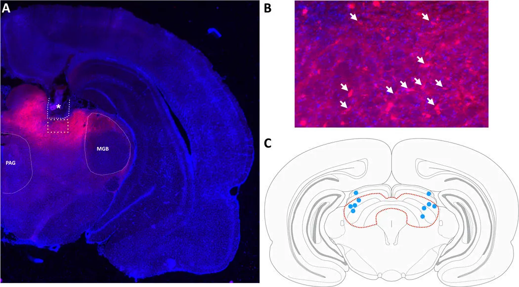
Figure A: * indicates the implantation location of the fiber optic probe and cannula;
Figure B: Arrows indicate mCherry-positive (expressing chr2) neuronal cells;
Figure C: Brain atlas of the deep and middle layers of the colliculus in the coronal plane – Safwan K. Hyder, et al., Optogenetic activation of the superior colliculus attenuates spontaneous seizures in the pilocarpine model of temporal lobe epilepsy
Case 2
——Erik Naylor, et al., Integration of Optogenetic Stimulation with Neuronal and Neurotransmitter Recordings - Kindling Using Optogenetics
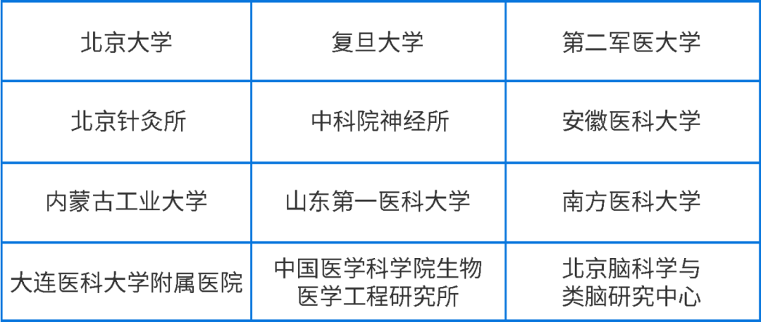
Partial user list